Manufacturer > Boston Scientific > Devices > OPTICROSS™ 35

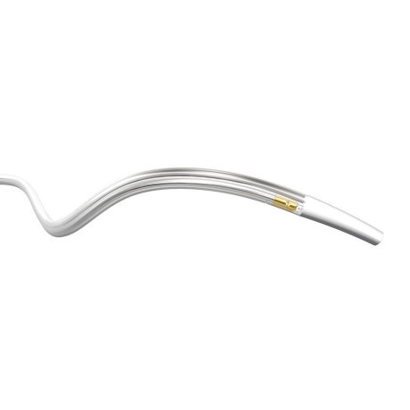
OPTICROSS™ 35

Device-Type

Intravascular Ultrasound

Targated Speciality

Cardiovascular

Manufacturer

Boston Scientific


The OptiCross 35 peripheral imaging catheter is the latest addition to Boston Scientific’s IVUS portfolio. The catheter’s unique design provides exceptional image quality leading to more confident detection and treatment of venous disease.

Features and Benefits

Unique Design

  • OptiCross 35 uses a first of it’s kind rotating transducer to produce a TRUE 360 degree view.
  • 1800 RPM Rotation Speed

High Image Quality

  • The images produced by the OptiCross 35 catheter deliver excellent clarity and a large depth of field.
  • 70 mm Target Imaging Diameter
  • 15 MHz Target Operating Frequency

Optimal Outcomes

  • Clear IVUS images help refine treatment pathway to provide a reliable plan and improve patient outcomes.

Recommended Compatible Devices

Necessary Equipment

The following are required for use of the OptiCross 35 Peripheral Catheter:

Use Case Examples

This catheter is intended for ultrasound examination of peripheral pathology only. Intravascular ultrasound imaging is indicated in patients who are candidates for transluminal interventional procedures.

Safety informations

CONTRAINDICATIONS

OptiCross 35 15 MHz Catheter: This product is contraindicated in the presence of conditions which create unacceptable risk during catheterization. This device is not to be used in the coronary arteries. MDU5 PLUS Sterile Bag: None known.


WARNINGS

  • Do not pinch, crush, kink or sharply bend the catheter at any time. This can cause poor catheter performance, vessel injury or patient complications. An insertion angle greater than 45° is considered excessive and can lead to device kinking which can cause difficult advancing/retracting the telescope shaft and poor image quality.
  • DO NOT advance the catheter if resistance is encountered. The catheter should never be forcibly inserted into lumens narrower than the catheter body or forced through a tight stenosis.
  • When advancing or retracting the catheter through a stented vessel, the catheter tip may interact with the stent, resulting in entrapment of catheter/guidewire, catheter tip separation, and/or stent dislocation.
  • If resistance is met upon withdrawal of the catheter, assess the cause of resistance using fluoroscopy, and if necessary, remove the entire system as a unit.
  • Do not advance the catheter along a guidewire that may be passing between one or more stent struts. This could result in entrapment of catheter/guidewire, catheter tip separation and/or stent dislocation. Use caution when introducing or removing the catheter from a stented vessel.


PRECAUTIONS

Do not advance the imaging core if resistance is encountered

Potential adverse events

The risks and discomforts involved in vascular imaging include those associated with all catheterization procedures. These risks or discomforts may occur at any time with varying frequency or severity. Additionally, these complications may necessitate additional medical treatment including surgical intervention and, in rare instances, result in death. Complications may include:

  • Allergic reaction (device, contrast or other)
  • Arrhythmia
  • Device entrapment requiring surgical intervention
  • Embolism (air, foreign body, tissue or thrombus)
  • Hemorrhage/Hematoma
  • Hepatic dysfunction
  • Hypotension/hypertension
  • Infection/sepsis
  • Ischemia
  • Renal insufficiency/failure
  • Stroke/cerebrovascular accident/transient ischemic attack
  • Thrombosis/thrombus
  • Vasospasm
  • Vessel injury (including perforation, trauma, rupture, dissection, and pseudoaneurysm)
  • Vessel occluion

Device Documents

Related Device SKUs

* Select 2 or more (upto 4) SKUs to compare.
SKU Name Device SKUs Model Transducer Frequency Typical Use Maximum Diameter Penetration Prep Location Catheter Telescoping Length Sled Pullback Length Distance from Transducer to Tip Guidewire Lumen Length Guidewire Compatibility Sheath Compatibility (with max wire) Crossing Profile Entry Profile Working Length
OPTICROSS 35
H7493932800350 15 MHz Common & External Iliac, IVC, Femoral 70 mm Proximal 25 cm 10 cm 1.0 cm 105 cm ≤ 0.035" 8 F 8 F 6 F 105 cm
device-thumbnail
OPTICROSS 35
  • Model : H7493932800350
  • Procedure : -
  • Transducer Frequency : 15 MHz
  • Typical Use : Common & External Iliac, IVC, Femoral
  • Maximum Diameter Penetration : 70 mm
  • Prep Location : Proximal
  • Catheter Telescoping Length : 25 cm
  • Sled Pullback Length : 10 cm
  • Distance from Transducer to Tip : 1.0 cm
  • Guidewire Lumen Length : 105 cm
  • Guidewire Compatibility : ≤ 0.035"
  • Sheath Compatibility (with max wire) : 8 F
  • Crossing Profile : 8 F
  • Entry Profile : 6 F
  • Working Length : 105 cm

Questions & Answers


Don't see the answer you're looking for ?