Device-Type
Intravascular Ultrasound
Manufacturer
Boston Scientific
iLAB™ Ultrasound Imaging System and ULTRA ICE™ PLUS Ultrasound Imaging Catheter for Intracardiac Imaging
ICE provides the combination of real-time imaging and soft tissue visualization that cannot be duplicated by fluoroscopy, pre-operative imaging (CT or MR), or electroanatomic mapping. Not only can you identify anatomical structures, you can visualize where devices are relative to those structures.
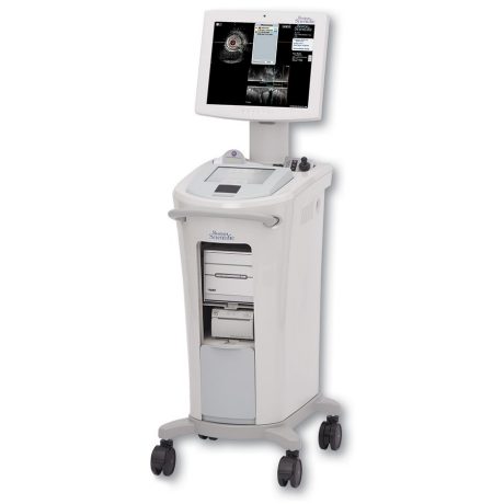
iLAB Ultrasound Imaging System
iLAB transformed visualization during EP procedures as the first intracardiac ultrasound system customized for the EP lab. It offers an easy user interface and automatic enhancement of ICE images. The Modular hardware design comes either installed or on a cart allowing flexibility to upgrade. The iLAB system is compatible with all Boston Scientific ultrasound catheters: Intracardiac (ICE), Intravascular (IVUS) and Peripheral (PI).
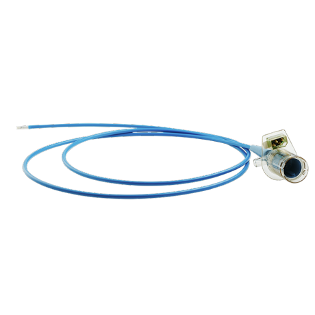
ULTRA ICE PLUS Catheter
The Boston Scientific ULTRA ICE PLUS catheter is designed to provide precise, real-time visualization of both intracardiac anatomy and devices positioned within the heart. Not only does it help you in identifying anatomical structures, it also helps you in visualizing where your devices are relative to those structures.
Device Illustration

Unique Vision: 360° Views and Detailed Near-Field Resolution
The ULTRA ICE PLUS Catheter generates a cross-sectional and panoramic 360° image perpendicular to the catheter, with the tip as a central reference point. This allows the user to visualize structures (such as the fossa) directly adjacent to the catheter tip and still see a detailed cross-section of the entire septum.
Image Comparison

Features and Benefits
The ULTRA ICE PLUS catheter is indicated for enhanced visualization of cardiac structures and is designed to optimize performance when imaging by enabling users to position the catheter directly adjacent to areas of interest, like the proximal coronary sinus, fossa ovalis, or pulmonary veins. Its 360 degree, forward looking visualization field provides greater near-field resolution than phased array catheters, without the need to manually move the catheter away from a target area to adjust focal distance.
Know Where You Are

ULTRA ICE PLUS catheter positioned in the right atrium, adjacent to the fossa ovalis, visualizing the structures critical to successful transseptal puncture: the septum, aorta, needle position, tenting, and the LAFW.
See What You Want to Avoid

Notice the patient’s reduced Left Atrium, the tenting of the septum and its relationship to the LAFW. The corresponding fluoroscopic image may suggest that puncture has already occurred. However, the ULTRA ICE PLUS image shows that this is not the case and guides the physician to redirect the needle in a puncture angle away from the LAFW.
Crossing to the Septum to Help Guiding Left Sided Procedures

A key application for the ULTRA ICE PLUS catheter involves crossing the septum and then monitoring and helping to guide left-sided procedures. In this setting, ULTRA ICE PLUS catheter is designed to allow the user to:
- Visualize left atrial anatomy
- Confirm catheter location relative to the anatomy
- Verify tip-to-tissue contact
- Identify location of the esophagus relative to the ablation catheter
- Characterize acute lesion morphology: swelling, dimpling, and crater formation
- Monitor for any early signs of thrombus formation, stenosis, or pericardial effusion